
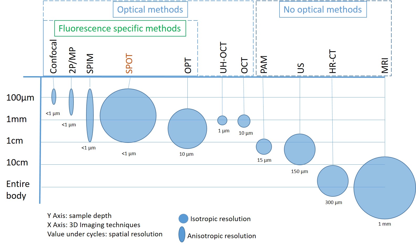
3D imaging of biological tissues is getting more and more attention in the last decades, due to the great amount of information that can be derived from the full stack of images. There are several 3D Imaging techniques, that can be divided into optical and non optical methods. Non optical methods like Magnetic resonance (MRI), Photoacoustic microscopy (PAM), Ultrasound (US) and High-resolution computed tomography (HRCT) are very popular in live specimen 3D imaging due to their capacity of generating contrast between different types of tissues. Their capacity of large sample imaging and isotropic resolution make them ideal for diagnostics but the low resolution images cannot be used for research in cellular and subcellular level.
The optical methods in general terms are ideal for high resolution imaging but the size of sample that can be imaged is significantly lower. Optical tomography methods like Optical Coherence Tomography (OCT), Ultra-High resolution Optical Coherence Tomography (UH-OCT) and white light absorbance Optical Projection Tomography (OPT) are providing isotropic 3D images with cellular resolution but with no specific information.
Confocal, Multiphoton and Selective Plane Fluorescence microscopy are the ideal techniques for high resolution cellular and subcellular resolution 3D images, but they present very poor axial resolution, especially in large tissues due to light diffraction from the inner sample. The only optical platform that can provide 3D images of mid size samples (up to 20mm), with cellular true 360º isotropic resolution, is our QLS-scope microscope, thanks to our patented Statistical Optical Projection Tomography (SPOT) technology.
For more information on our technology, please contact with us: info@planelight.net