Molecular Imaging of Collagen Destruction of the Spine
” ABSTRACT: As the leading cause of disability worldwide, low back pain is commonly
caused by biomechanical and catabolic disruptions to key structures of the spine, such as
intervertebral discs and facet joints. To date, accurate, noninvasive detection of
microdestruction within these tissues remains an elusive goal. Here, we report an in vivo
imaging approach based on a collagen hybridizing peptide (CHP) that specifically targets
disruption to the extracellular matrix architecture at the molecular scalethe denatured
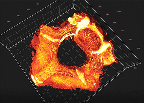
collagen molecules. Utilizing fluorescently labeled CHPs, live animal imaging, and light
sheet fluorescence microscopy, we mapped collagen destruction in the lumbar spines in 3D,
revealing that under normal conditions collagen destruction was localized to load-bearing
anatomical structures including annulus fibrosus of the disc and the facet joints, where
aging, tensile force (hindlimb suspension), and disc degeneration (needle puncture) escalated the CHP-binding in specific
mouse models. We showed that targeting denatured collagen molecules allowed for an accurate, quantifiable interrogation of
the structural integrity of these spinal matrixes with a greater sensitivity than anatomical imaging and histology. Finally, we
demonstrated CHP’s binding to degenerated human discs, suggesting exciting potentials for applying CHP for diagnosing,
monitoring, and treating various spinal disorders, including intervertebral disc degeneration, facet joint osteoarthritis, and
ankylosing spondylitis.
KEYWORDS: spinal disorder, intervertebral disc degeneration, facet joint, microdestruction, collagen triple helix “
Total article:
Molecular Imaging of Collagen Destruction of the Spine | ACS Nano
